Peri-implant disease is unarguably one of the most significant risks
associated with implants. It is a multifactorial disease, which if not
diagnosed at early stage, can ultimately lead to failure of the implant.
WHAT IS PERI-IMPLANT DISEASE?
Peri-implant disease is a condition that affects the tissues surrounding a functional implant; it includes both peri-implant mucositis and peri-implantitis.
Peri-implant mucositis can be defined as ‘reversible inflammatory reactions in the soft tissues surrounding a functioning implant.
Peri-implantitis is characterised by ‘inflammatory reactions with loss of supporting bone in the tissues surrounding a functioning implant.


Diagnosis of peri-implant disease relies on
crude parameters commonly used for the diagnosis of periodontal disease.

WHAT IS PERI-IMPLANT DISEASE?
Peri-implant disease is a condition that affects the tissues surrounding a functional implant; it includes both peri-implant mucositis and peri-implantitis.
Peri-implant mucositis can be defined as ‘reversible inflammatory reactions in the soft tissues surrounding a functioning implant.
Peri-implantitis is characterised by ‘inflammatory reactions with loss of supporting bone in the tissues surrounding a functioning implant.
Peri-implantitis yields many features in common with chronic
periodontitis.
Both involve alveolar bone loss.
However, there is a
zone of connective tissues which is attached to the root surface in
periodontitis.
In contrast, connective tissue does not attach directly onto
implants and there is no periodontal ligament. Therefore, the inflammatory lesion
in peri-implantitis extends closer to the bone surface, which can be associated
with a faster rate of progression and more aggressive consequences.


AETIOLOGY AND RISK FACTORS
Gram-negative
anaerobic bacteria, such as Porphyromonas
gingivalis, Prevotella intermedia
and Actinobacillus actinomycetemcomitans. Bacterial flora that
are associated with periodontitis and peri-implantitis are found to be similar
Implants in partially
dentate patients appear to be at a greater risk of peri-implantitis than
implants in fully edentulous patients. Natural teeth serve
as reservoirs for periodontal pathogens from which colonisation of the implant
sites occurs.
patient-related risk
factors include: inadequate oral hygiene, smoking, parafunctional habits and underlying
systemic conditions such diabetes.
occlusal overload will play an important role implant failure by resulting in progressive bone
loss around the implant.
Iatrogenic factors
such as lack of primary stability, poorly positioned implants, premature
loading during the healing period and poorly fitting abutments or restorations.
DIAGNOSIS
Swelling and redness of the peri-implant marginal
tissues and plaque/calculus accumulation are important signs.
Bleeding on probing and suppuration are clear
indications of disease.
successful implants generally allow a probe penetration
of approximately 3-4mm in the peri-implant sulcus.
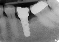
Adequate baseline radiographs determine the peri-implant
bone status as well as the marginal bone level. These can then be compared to
future radiographs to determine if additional bone loss, beyond ‘normal’ has
occurred. Progressive bone loss is a definite indicator of
peri-implantitis.
Implant mobility is an insensitive measure in
detecting early implant failure
More advanced peri-implantitis is characterised
by mobility of the fixture, indicating failure of osseo-integration.


MANAGEMENT
When the main aetiological factor is bacterial
infection, the first phase of treatment involves the control of acute infection
and the reduction of inflammation. This involves the removal of plaque deposits
and improved patient compliance with oral hygiene until a healthy peri-implant
site is established.
The implants that are affected with
peri-implantitis are contaminated with soft tissue cells, microorganisms and
microbial by-products. The defect must be debrided. Prophy jet and the use of a
high pressure air powder abrasive has been advocated, as this removes the
microbial deposits, does not alter the surface topography and has no adverse
effect on cell adhesion.
contact with a supersaturated solution of citric
acid have been used for the preparation of the implant surfaces.
Soft tissue
laser irradiation has also been used .
systemic administration of antibiotics that
specifically target gram-negative anaerobic organisms has shown an alteration
in the microbial composition and a sustained clinical improvement. A local delivery device with fibers containing polymeric
tetracycline has been tried and this resulted in significantly lower total
anaerobic count.
If vertical 1 to 2-wall defects (< 3mm) are
found, then the resective surgery may be used to reduce the pockets, to
smoothen the rough implant surfaces, to correct the osseous architecture and to
increase the area of the keratinized gingiva.
Various bone grafting techniques and materials
and guided bone regeneration, have been successfully used for the regeneration
in 3-wall or circumferential defects.
Porous titanium granules have also
recently been advocated to try and treat advanced peri-implant osseous defects
When biochemical forces are considered as the
main aetiological factors, occlusal equilibration i.e. improvement of
the implant number and position and
changes in the prosthetic design, can arrest progression.
No comments:
Post a Comment