
Saturday, December 14, 2013
Tuesday, December 10, 2013
Wednesday, June 26, 2013
Radiation-Induced Xerostomia and its treatment
Radiation-induced xerostomia is caused by the direct damaging effects of radiation on both major and minor salivary gland structures located in the radiation path.
Glandular tissue in general is very sensitive to radiation. Following radiation therapy, the mouth becomes dry as a result of the loss of salivary gland acini. The skin becomes dry as well because of loss of sweat and sebaceous glands.

Ductal epithelium is somewhat radiation resistant.

Glandular tissue in general is very sensitive to radiation. Following radiation therapy, the mouth becomes dry as a result of the loss of salivary gland acini. The skin becomes dry as well because of loss of sweat and sebaceous glands.
Ductal epithelium is somewhat radiation resistant.
Because most radiation ports leave some areas of mucosa untouched, there is an opportunity to stimulate the remaining glands to overproduce.
Although they improve mouth moisture in only about 70% of irradiated patients,
pilocarpine (Salagen, MGI Pharma), 5 mg by mouth three times daily, or
cevimeline (Evoxac, Daiichi Sankyo), 30 mg by mouth three times daily, often improves eating, speaking, and swallowing functions.
It must be given with caution to individuals with heart diseases associated with brachycardia, heart block, or medications such as beta blockers that may slow heart rate or conduction.
Additionally, sports water bottles are used by many individuals, and Evian atomized water spray has been found to be beneficial to many.
Radiation Caries and its management
Radiation caries results from xerostomia, which permits cariogenic bacteria to proliferate unopposed by the usual lysosomes and IgA immunoglobulins in saliva and causes the loss of the saliva’s natural buffering capacity.
Caries in nonirradiated individuals occurs in pits, in fissures, and interproximally. It is also chalky and soft from dissolved tooth structure.
Radiation caries, by contrast, is hard and black. It occurs at the gingival margin, cusp tips, incisal surfaces, or throughout the tooth.

Monday, June 17, 2013
Factors to be Considered when preparing a root canal
With the invention of nickel titanium instruments came the additional question of, “How wide should I make the taper of the preparation?” This essentially split clinicians into two schools of thought.
1. Those who prepare the apex to large apical sizes, such as 40 and above but use narrow tapers (4%).
2. Those who prefer less apical preparation size 20 or size 25 but use a wide taper (6% and above).

Thursday, May 23, 2013
Indications and potential limitations of Gates Glidden drills (GGDs) in endodontics
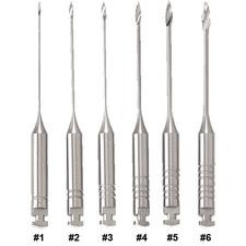
Gates Glidden Drill Basics
GGDs have been used for decades to shape the orifice and canal above the point of first curvature in endodontic procedures.
Traditionally they come in lengths of 28, 32 and 38 mm and in sizes #1-6, with size #1 being the smallest in diameter and #6 the largest.
Saturday, May 18, 2013
Gingival Mask for gingival recession patients
A Gingival mask is a removable appliance for patients who have lost tissue due to trauma, perio surgery, or recession, and when methods such as surgery or other regenerative procedures are considered unpredictable, impossible, or have failed.


PERI-IMPLANT DISEASE, A GROWING PROBLEM
Peri-implant disease is unarguably one of the most significant risks
associated with implants. It is a multifactorial disease, which if not
diagnosed at early stage, can ultimately lead to failure of the implant.
Thursday, February 21, 2013


Sunday, February 17, 2013
Permar’s Oral Embryology and Microscopic Anatomy 10th Edition PDF - e book

Description
Now in its Tenth Edition, Permar’s Oral Embryology and Microscopic Anatomy continues to provide comprehensive, yet concise coverage of embryology and histology for dental hygiene anddental assisting professions. It can also be used as an introductory text for dental students. This text begins with the basics of general histology, progresses through the development of the human embryo and fetus, and concludes with a focus on the development of the face and oral cavity. New to this edition are over 40 additional illustrations, including four-color micrographs. High-quality images of microscopic embryonic development and oral anatomy help students identify histologic structures. A new chapter regarding salivary glands includes information about remineralization, demineralization, fluoride, bacterial diseases, and HIV. Clinical aspects of oral tissue are covered to help readers expand their knowledge from basic to clinical sciences and apply fundamental principles. Suggested readings help readers find additional resources.
DOWNLOAD HERE DUDES ;)
Surgical Anatomy of the Face 2nd Edition PDF - free e book

Description
Thoroughly updated to reflect the latest refinements in operative technique, this full-color atlas provides a surgeon’s-eye view of the anatomic structures and relationships encountered during all facial surgical procedures. It features more than 100 drawings by Dr. Makielski, a head and neck surgeon, and more than 100 photographs.
This Second Edition’s brand-new chapter on embryology emphasizes congenital anomalies such as clefts and dermoid cysts. New illustrations show the surgical anatomy of endoscopic approaches and recently developed procedures, including the SOOF lift. This edition also includes more detail on the osteocutaneous and retaining ligaments and the supporting ligaments and tendons of the orbit.
click here to go for the book ;)


Management Of Endodontic Emergencies
MANAGEMENT
OF PAINFUL IRREVERSIBLE PULPITIS
Because
the pain is the result of inflammation, primarily in the coronal pulp, removal
of the inflamed tissue will usually reduce the pain.
Without
Acute Apical Periodontitis
Complete
cleaning and shaping of the root canals is the preferred treatment if time permits.
With limited time, most pulpal tissue is
extirpated with a broach (partial pulpectomy) in single-rooted teeth.In molars,
a partial pulpectomy is performed on the largest
canals (palatal or distal root). Pulpotomy is usually effective in molars when minimal time
is available.
An old
but still popular idea is that chemical medicaments sealed in chambers help
control or prevent additional pain; this is not true. A dry cotton pellet alone
is as effective in relieving pain as a pellet moistened with camphorated
mono-chlorophenol (CMCP), formocresol, Cresatin,eugenol, or saline. Therefore,
after irrigation of the chamber or canals with sodium hypochlorite a dry cotton pellet is
placed and
the access is sealed temporarily. A mild analgesic may be prescribed for patients
with irreversible pulpitis. Antibiotics, however, are definitely not indicated.
Thursday, January 24, 2013


Tuesday, January 22, 2013


Sunday, January 20, 2013


Subscribe to:
Comments (Atom)